
In 1963, Dr. Lofgreen wrote an article for the Western Livestock Journal titled: “Net energy – the new way to reckon rations.”
He wrote that to further the daily use of the California Net Energy System and to gain common use of the net energy terms as standard feedlot lexicon.
A proper reference to Dr. Lofgreen in any manner possible ensures that his legacy endures and to note the importance of his and Dr. Garrett’s timeless work.
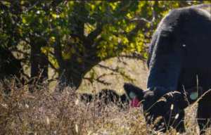
Now, let’s discuss digital dermatitis/hairy heel wart/hairy wart/papillomatous digital dermatitis/bovine digital dermatitis/strawberry.
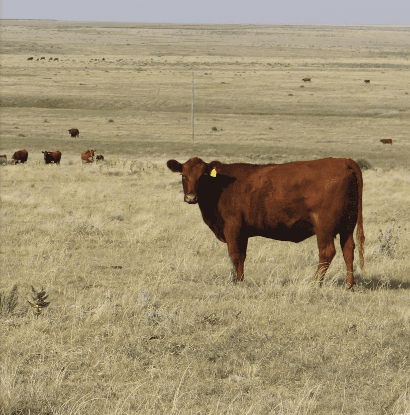
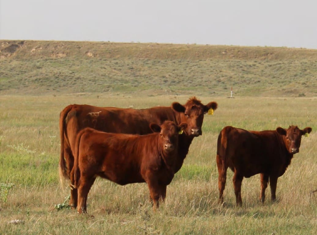
Feedyards, cattle owners, and professional advisors have dealt with the management and economics of an increase in feedlot cattle lameness.
We have made great strides in identifying, treating, and now proactive management of digital dermatitis (DD).
Digital dermatitis robs cattle of retained energy, decreases daily gain, and lowers overall performance. An example of loss in performance from Kulow et al. (2017) was a 0.31 lb/d lower ADG and 12 lb lighter hot carcass weight.
Regression data from Döpfer et al. (2018) showed that feedlot heifers with M4 lesions lost 0.34 lb/d of carcass weight compared with cattle without DD.
The decrease in body weight gain and the management of lame cattle, whether it is treating individuals or housing them in designated pens, is costly and adds another level of daily management at feedyards.
As has been described by many, DD was first reported in 1974 in Italy (Cheli and Mortellaro, 1974).

The classification of the types of DD lesions was described by Döpfer et al. (1997). Those authors classified the lesions as M-stages with the M standing for Mortellaro.
It is important to understand these M-stages. To further review the DD terms, here are the M-stages defined by Döpfer:
M0-Normal digital skin without signs of DD.
M1-Early, small circumscribed red to gray epithelial defect of less than 2 cm in diameter that precedes the acute stages of DD (M2). In addition, M1 stages can appear between acute episodes of DD lesions or within the margins of a chronic M4 lesion as an intermediate stage.
M2-Acute, active ulcerative (bright red) or granulomatous (red-gray) digital skin alteration, >2 cm in diameter, commonly found along the coronary band in addition to around the dew claws, in wall cracks, and occasionally as a sole defect.
M3-Healing stage within 1 to 2 days after topical therapy, where the acute DD lesion has covered itself with a firm scab-like material.
M4-Late chronic lesions that may be dyskeratotic (mostly thickened epithelium) or proliferative or both. The proliferations may be filamentous, scab-like or mass proliferations.
M4.1-The additional stage refers to the chronically affected foot that displays the M4 stage in addition to the M1 stage.
The primary points to be made from the above classification is that cattle develop small, gray, scaly or flaky lesions that develop into M2
lesions.
The M2 lesions typically cause cattle to express signs of lameness.
Cattle with DD may stand with a raised hoof and not wanting to stretch the hoof out, keeping it bent distally with the point of the claw pointing down to prevent any skin movement on the heel where there is an active lesion.
They may shake their affected hoof. An observant feedyard owner that is well schooled on DD termed the phrase “heel wart dance”, where cattle shift weight from hoof to hoof to relieve pressure and discomfort.
Other characteristics of lame cattle with DD are cattle that lay flat on their side to get weight off of their hooves or cattle that lay at the back of a pen or on a bed-pack for longer than normal intervals.
These cattle have been observed to have more tag covering the majority of one side of their body.
Cattle with DD commonly have less rumen fill and go to the bunk less frequently, potentially over consuming ration resulting in digestive disturbances.
A colleague observed a higher risk of bloat in cattle with DD due to erratic bunk attendance. Those are subtle observations.
They are important and commonly used to determine if a pen of cattle should walk through a foot bath.
The goal is to make sure that if there are any observed DD lesions, that pen is moved through a foot bath to prevent the development of advanced lesions (M2 or M4).
Dairy research (Robcis et al., 2023) reported that it was more efficient time management to use foot baths versus pulling and treating dairy cattle for DD.
Experience at feedyards confirms that to be true. In addition, feedyards may not have chutes equipped for handling hooves safely, further emphasizing that foot baths are safe, effective, and efficient in the intervention for DD.
Most feedyards are executing this well. The feedyards that have a prevalence of M2 and M4 lesions need to improve their processes.
Foot bath requirements include:
1) long enough for all hooves to be submerged twice
2) deep enough to cover the dew claws
3) in a location where cattle move in a normal gait and safely
4) re-charge the foot bath after each pen
5) drain and replace the foot bath solution when it is dirty, which may be after 100 head in small foot baths or after 1000 or more head in large foot baths.
It is critical to ensure that there is active ingredient in the solution.

Researchers from University of Wisconsin, Iowa State University, USDA Bacterial Diseases of Livestock Research Unit in Ames, and University of Calgary have provided us with insight into the etiology and polybacterial characteristics of DD.
A significant education has been provided by feedyards in western Iowa that have learned to manage the disease resulting in a decrease in M2 and M4 lesions.
A recent paper by Caddey and De Buck (2021) described the microbial community of these lesions.
Their meta-analysis showed that Treponema, Mycoplasma, Porphyromonas, and Fusobacterium have roles in DD.
Of those species, the Treponemes have the largest relative abundance in DD lesions.
Treponemes cause multiple bacterial infections in different species including humans and including relapsing skin infections (Antal et al., 2002).
In a review by Palmer and O’Connell (2015), the authors describe hoof conformation playing a role in risk level of cattle developing DD.
The height of the heel and the interdigital space have been shown to be a risk for developing DD such that lower heel height and narrower interdigital gap have increased risk.
Observations of mine and others of cattle with heel wart agree with the report of Palmer and O’Connell (2015) where claws wear abnormally resulting in higher heel height because cattle walk on the front of the claws wearing them down resulting in abnormal growth and less wear on the heel.
I classify cattle with abnormal hoof growth and DD to be cattle that were missed for treatment or intervention.
In the past, the common thought was that DD manifested after 120 days on feed.
We have now learned that lesions occur at any time during the feeding period. Expression of the disease is more commonly observed at heavier weights.
An example of this occurred in an experiment conducted in Iowa (Döpfer et al., 2018) where the incidence was less than 3-5% through 187 days on feed and from that point on, as cattle were fed between 320 and 380 days, incidence moved between 22 and 27%.
My thoughts on why we observe heavier cattle to more commonly have the disease is that it is a factor of the pressure on the hoof and lesion causing pain when cattle are heavier.
There is a picture of a lesion on page 156 in the Wilson-Welder et al. (2015) review that was described as “a less typical lesion of DD (red circle).”

That type of lesion is common in the interdigital space on the front of the hoof (Figure 1). These lesions are easily observed on the front hooves but commonly are not associated with lameness from my observations.
However, when front hooves are infected with D, weight loss is common due to lower bunk attendance and feed intake.
The length of time for DD to advance from an M1 lesion to an M2 takes time.
Refer to the M-scores from Döpfer and colleagues (1997). The progression of M1 to M2 is likely days.
The window for prevention is short. During the summer of 2024, in conjunction between Iowa Beef Council, Dordt University, myself, and colleagues (Mr. Evan Vermeer, Dr. Wes Gentry, and Dr. Dan Thomson), an observational experiment was conducted.
The objective was to identify 20 steers with active DD and treat them with 1 of 5 treatments.
The treatments were applied directly to the lesion while the steer was restrained in a chute and the affected hoof positioned for observation and treatment of the lesion.
The treatments were the exact solution as used in foot baths and the positive control was lincomycin.
The treatments were:
1. Lincomycin
2. Iowa formula (salt, copper sulfate, and soap)
3. Formalin
4. Benzoic acid (Dragonhyde)
5. Sodium bisulfite (BeefUp)
As mentioned, this was observational. The steers were weighed on the day of the initial treatment and housed in a pen in a bedding barn.
Subsequent treatments and weights occurred in 14, 15, and 20 day increments.
Each treatment showed efficacy based on observations of lameness and body weight gain. Lincomycin, formalin, and sodium bisulfite had the most improved lesions.
The benzoic acid product (Dragonhyde) coated the hoof very well though it also coated adjacent surfaces as well.
Keep in mind that this experiment had minimal replication.
The objective was to understand acutely how typical foot bath solutions affected DD lesions.


Figures 2 and 3 show the change in an M2 lesion over a 14-day period after being treated with the Iowa formula.


Figures 4 and 5 show the change over the same period in an M2 lesion after being treated with formalin.


Figures 6 and 7 show the change in a DD lesion after being treated with benzoic acid (Dragonhyde).


Figures 8 and 9 show the change in a DD lesion after being treated with sodium bisulfite (BeefUp). The conclusion was that doing anything to disrupt the lesion’s biofilm was beneficial.
These cattle (primarily M2 lesions) gained 3.27 lb/d from 1385 to 1545 lb during this period.
We did not have a comparison of gain for cattle during that period that did not have DD.
More university and industry evaluations of foot bath solutions need to occur. Many products are used.
There are products like formalin that are effective but have safety concerns and a need for PPE to be used.
Concentrations of products vary. Based on conversations with feedyards and industry professionals, there are preferred concentrations.
There are a lot of comments that may or may not be based on any good information, let alone any hard data.
Copper sulfate and zinc sulfate have efficacy, but there are issues with what to do with the spent foot bath solution.
There are excellent veterinarians working to help identify DD treatments.
Professional hoof trimmers and feedyards are trimming hooves of lame feedlot cattle when necessary. From those interventions, advancements in getting cattle comfortable, allowing more weight to be gained, are occurring.
Dr. Dan Thomson brought sodium bisulfite to our attention, which led Dr. Engelken at Iowa State University to conduct an experiment with it as a pen surface treatment. Its efficacy is being evaluated at other yards now.
Our experience is that multiple products have efficacy in getting cattle to exhibit less lameness which confirms previous data from Teixeira et al. (2010), presumably indicating less pain.
This improves DMI post treatment which increases body weight. The improved locomotion and comfort of cattle is noticeable within hours to days following foot bathing.

Diets formulated by professionals considering industry norms (Samuelson et al., 2015) and at or exceeding requirements (NRC, 1996) are fundamental.
Feedyards, cattle feeders, and professionals that have experience with a wide range of feedstuffs and diets understand that there are multiple variables contributing to the disease and that there are important nutritional characteristics to understand.
There are people in the feed business that claim specific feedstuffs cause the disease or that a specific as fed percent inclusion will lead to DD.
Those that have been involved over a duration of years and a range of diets understand that DD is not caused by an ingredient.
Dogma based on limited observations or perpetuated by large personalities is folly.
A feedyard in western Iowa in the Loess Hills had finishing cattle and dairy replacement heifers fed the same ingredients in diets formulated for the different production goals.
The finishing cattle had DD, whereas the dairy heifers did not express DD.
There are other diet-related observations that do point to a combination of ingredients and/or cattle movement or transport that may influence an environment where DD is more prevalent.
These are important observations but are not to be considered causative but part of a complex microbiome at each feedyard.
There may be specific nutrients that become more concentrated in tissue that are used by specific bacteria to grow. These are unproven theories at this point.
Work that Zinpro has sponsored (Kulow et al., 2017 and Döpfer et al., 2018) shows an improvement in carcass weight and less M2 and
M4 lesions.
The feedlot industry adopted the use of more bioavailable trace minerals long before DD was a widespread issue.
Diamond V NutriTek (NaturSafe for beef cattle) has shown efficacy (Anklam et al., 2025).
Biotin has been shown to decrease DD in dairy cattle over a 12-month period (Hochstetter et al., 1998).
The conundrum that we have is that all products add up to significant cost. Nutritional intervention is a cost to all cattle.
The benefit to heel wart control may only affect 25% of the cattle with DD.
At times, the cost of the intervention to the pen or feedyard as a whole is more than the benefit.
It is implicit that if a feedyard trims hooves, the individual doing the trimming is well trained by a professional hoof trimmer.
For example, pictures sent to knowledgeable veterinarians or hoof trimmers can be used to give guidance to improve technique and outcomes.
There is great knowledge and experience in that arena. Be mindful of clean instruments.
Wells and colleagues (1999) pointed out that unclean trimming tools can spread the disease.
This depends on the severity and number of affected cattle.
The typical protocol is to move cattle through a foot bath multiple days per week if the feedyard is behind treating for DD and have greater than 2% lame cattle in the pen.
The 2% threshold is arbitrary. If there are cattle with DD in the pen, a foot bath should be considered.
Consecutive versus every other day? My preference is consecutive days.
That is an aggressive approach to head off the problem. Many feedyards and advisers use that approach.
Out of precaution, the concentration of the active ingredient in the foot bath needs to be correct.
There are logistical and weather-related variables that dictate which is best at every feedyard.

We need to take a step back and assess production systems and consider previous management and environment.
Transport, handling, and facility hygiene need to be monitored so that we can correctly understand where DD originates.
There are bedding barns that are prone to have cattle with DD. Conversely, there are bedding barns that do not have cattle that express signs of DD.
There is unproven dogma related to feedstuffs. There are feedyards with basic vitamin and mineral nutrition that have no issues with DD.
The experience that we have gained is that feedyards that have had DD will not necessarily continue to have cattle that are infected with it.
It occurs in all pen designs. It appears in most breeds. It appears in most feeder cattle weight classes.
Appreciation is due to cattle feeders in western Iowa, most specifically Sioux County, who have helped teach the industry what DD is and how to manage it.
There are active companies and industry professionals searching for products and management strategies to lessen the effect of DD and ultimately to lead us into a prevention of the disease.
For now, diligent identification of DD and proactive management practices are proven to lessen the advancement of lesions.
New tools such as real-time DD detection apps such as developed by Dwivedi et al. (2025) should enable us to have the insight and eye for identification such as Dr. Dörte Döpfer and other highly trained personnel.
Critical contact points include:
• If a feedyard or pen has a history of DD, foot bath early
• Pen and trailer hygiene
• Cattle handling
• Proper nutrition
References
• Anklam, K., M. Aviles, J. Buettner, S. Henschel, R. Sanchez, S. Ordaz, I. Yoon, J. Wheeler, G. Dawson, and D. Doepfer. 2025. Evaluation of
Saccharomyces cerevisiae fermentation product on the prevention of digital dermatitis using an experimental infection model in cattle. App.
Anim. Sci. 41:47-64. doi.org/10.15232/aas.2024-02567
• Antal, G.M., S.A. Lukehart, A. Z. Meheus. 2002. Review: The endemic treponematoses. Microbes and Infection. 4:83-94
• Caddey, B. and J. De Buck. 2021. Meta-analysis of bovine digital dermatitis microbiota reveals distinct microbial community structures
associated with lesions. Front. in Cell. and Inf. Micr. 11:1-12. doi:10.3389/fcimb.2021.685861
• Cheli, R. and C. Mortellaro. 1974. La dermatite digitale del dovino. In: 8th International Meeting on Diseases of Cattle, Milan, Italy. p.
208–213.
• Döpfer, D., A. Koopmans, F. A. Meijer, I. Szakall, Y. H. Schukken,W. Klee, R. B. Bosma, J. L. Cornelisse, A. vanAsten, and A. terHuurne.
1997. Histological and bacteriological evaluation of digital dermatitis in cattle, with special reference to spirochaetes and campylobacter
faecalis. Vet. Rec. 140(24):620–623 (Article). doi:10.1136/vr.140.24.620
• Döpfer. D. E.R. Loe, C.K. Larson, and M.E. Branine. 2018. Effects of feeding a novel amino acid-complexed trace mineral supplement
on productivity and digital dermatitis mitigation in growing-finishing heifers. J. Anim. Sci. 96:suppl 2, Page 231, doi.org/10.1093/jas/
sky073.428
• Dwivedi, A. M. Henige, K. Anklam, and D. Döpfer. 2025. Real-time digital dermatitis detection in dairy cows on Android and iOS apps
using computer vision techniques. Transl. Anim. Sci. 9:1-13. doi.org/10.1093/tas/txae168
• Hochstetter, T. 1998. Horn quality of the bovine hoof under the influence of biotin supplementation. DVM Inaugural Dissertation. Journal
#2176. Free University of Berlin, Germany.
• Kulow, M., P. Merkatoris, K.S. Anklam, J. Reiman, C. Larson, M. Branine, and D. Döpfer. 2017. Evaluation of the prevalence of digital
dermatitis and the effects of performance in beef feedlot cattle under organic trace mineral supplementation. J. Anim. Sci. 95.
doi:10.2527/jas2017.1512
• NRC. 1996. Nutrient requirements of beef cattle. 7th rev. ed. Natl. Acad. Press, Washington, DC.
• Palmer, M.A., and N.E. O’Connell. 2015. Digital dermatitis in dairy cows: a review of risk factors and potential sources of between-animal
variation in susceptibility. Animals. 5:512-535. doi:10.3390/ani5030369
• Robcis, R. A. Ferchiou, M. Berrada, and D. Raboisson. 2023. Management of digital dermatitis in dairy herds: optimization and time
allocation. Animals. 13:1-20. doi.org/10.3390/ani13121988
• Samuelson, K.L., M.E. Hubbert, M.L. Galyean, and C.A. Loest. 2016. Nutritional recommendations of feedlot consulting nutritionists:
The 2015 New Mexico and Texas Tech University survey. J. Anim. Sci. 94:2648-2663.
• Teixeira, A.G.V., V.S. Machado, L.S. Caixeta, R.V. Pereira, and R.C. Bicalho. 2010. Efficacy of formalin, Copper sulfate, and a commercial
footbath product in the control of digital dermatitis. J. Dairy. Sci. 93:3628-3634.
• Wells, S.J. and B.A Wagner. 1999. Papillomatous digital dermatitis risk factors in US dairy herds. Preventive Vet. Med. 38:11-24. doi.
org/10.1016/S0167-5877(98)00132-9
• Wilson-Welder, J.H., D.P. Alt, and J.E. Nally. 2015. The etiology of digital dermatitis in ruminants: recent perspectives. Vet. Med.: Res. &
Rep. 6:155-164. dx.doi.org/10.2147/VNRR.562072
 Erik Loe, PhD, has been a professional feedlot nutritionist since 2008. Prior to that, he was the SDSU Extension Feedlot Specialist and received graduate degrees from NDSU and K-State. He truly enjoys being at feedyards and working for them. He finds his colleagues professionally inspiring and challenging, a combination he values in line with one of his favorite quotes, from a previous National Security Advisor: “One always learns more from friendly critics than from uncritical friends.” Dr. Loe has many blessings, including being married to a wonderful lady and having two outstanding daughters.
Get all Doc Talk episodes straight to your email inbox!