
When talking about diseases affecting cattle and what we can do to support cattle health, it can often seem a bit like looking into a bowl of alphabet soup.
You have a few common players that most people know about: BVDV, IBR, BRSV, PI-3 and vaccines for these agents (many of which are MLVs).
Dairy cattle recently made IAV part of the cattle conversation.
The cow that stole Christmas a couple decades ago did so through a diagnosis of BSE, and there are several acronyms we all hope we never get to experience up close (namely, FMD).
Epizootic hemorrhagic disease virus (EHDV) and Bluetongue virus (BTV) are two separate but very similar viruses that can each cause issues in cattle from time to time.

As previously stated, these viruses are incredibly similar: they both belong to the same genus (Orbivirus) and often result in fairly similar signs/lesions in the primary species they affect.
EHDV most often affects white-tailed deer which are highly susceptible to infection; the most common species affected by BTV is sheep.
However, reports of confirmed disease due to these viruses extend to various species of deer and multiple different domesticated ruminants (goats, llamas, yaks, bison).
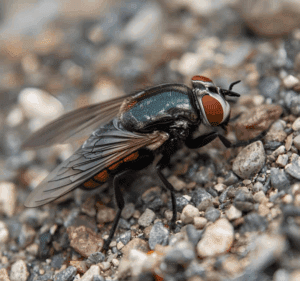
Multiple strains of both viruses exist (called serotypes) and both are spread via the Culicoides sp. of biting midges (aka, no-see-ums).
Other insects have been shown to have little to no role in virus transmission, so spread is essentially limited by the geographic areas where biting midges are present and the time of year where these insects are alive/active.
Deer affected by EHDV are often found dead; those that are not may be found breathing heavily with a high fever, weakness, and lameness.
The most striking changes often occur in the mouth and head/neck; the lining of the mouth often sloughs and tissues of the mouth/head/neck are often severely swollen.
Bloody saliva may be found coming from the mouth, and some animals may lose their hooves.
BTV in sheep can look similar but is generally not quite as severe. Affected sheep have a swollen face/muzzle, nasal discharge, and oral ulcers.
The tongue may be blue/swollen and sticking out of the mouth.
Death loss in white-tailed deer can be very high, particularly in captive/farmed deer operations with high population density.
The impact of BTV infections in sheep is variable and often depends on the strain as well as the level of existing immunity.
When we examine animals that have died due to these viruses, we typically see scattered hemorrhages throughout the carcass affecting many organs in addition to the previously discussed changes to the face/neck/ mouth.
Swelling due to edema and fluid within the lungs are also common; these changes all stem from damage to blood vessels caused by the viral infection.

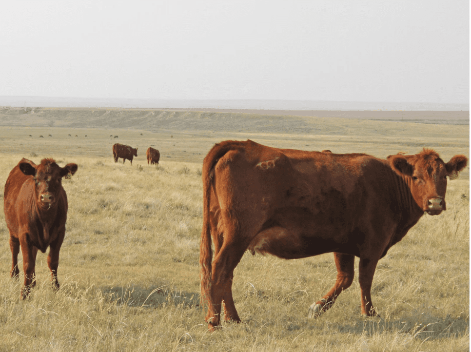
These viruses can become impactful in cattle when they are introduced into areas where the cattle herds have not previously been exposed.
Most infections of EHDV and BTV in cattle are thought to be either subclinical (not cause symptoms) or cause very mild/minor impacts on
the herd.
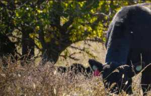
Changes are similar to but generally much, much milder than infections in whitetailed deer or sheep: swollen faces, ulcers in the mouth, excess salivation, and lameness are most common.
Death loss in cattle during outbreaks has reportedly been very low.
With that being said, reports during a recent EHDV outbreak in southern Iowa often described a significant number of cattle being found dead on pasture with severely swollen faces and sloughed tissue in the mouth.
Another potential impact in cattle is on fetal development in infected pregnant cows.
Both viruses can interrupt fetal development and result in abnormalities such as domed heads and small eyes; other more common viruses (like BVDV) can cause similar defects in fetuses.
Both of these viruses are fairly easy to detect in animals showing signs of disease; PCR tests have been developed for each of these agents that can be run using either whole blood from live cattle or spleen tissue collected at necropsy.
Brain tissue can also be used to detect each virus in cases of fetal defects. Tests for antibody to EHDV and BTV are also available to assess whether an animal has previously been exposed to either virus.
However, these viruses are similar enough to each other that antibody testing results can be difficult to interpret; a positive test for BTV antibody may actually be the result of previous EHDV infection, depending on the test.
Detection of these viruses and/or suspicion of disease due to them is reportable in many states.
Despite the widespread geographic distribution of the viruses and the biting midges that spread them, some countries require diagnostic testing prior to accepting animals or embryos.
Much of the routine testing we do for EHDV and BTV is for embryo export purposes to show that donors are negative at the time of collection; detection of either of these viruses in blood samples can potentially be disqualifying.
Consult your herd veterinarian if you have questions regarding embryo export, and talk through the export requirements and the logistics of meeting those requirements for each intended destination country.
Vaccination for these agents as a control measure is complicated: vaccines may be available or able to be produced in commonly affected areas of the world, but how well they work is highly dependent on the strain of virus present in the vaccine.
Most control measures involve reducing the exposure of animals to biting midges, but many of these recommendations just are not terribly feasible in large herds occupying large pastures.
Insect repellents may be effective in small areas.
Avoiding areas with heavy insect populations (low lying, damp ground) and times of high insect activity/feeding (dawn/dusk) may be helpful but difficult to execute.
 Drew Magstadt is a Clinical Associate Professor and has been a veterinary diagnostician for the past decade at the Iowa State Veterinary Diagnostic Laboratory. He grew up on a ranch in central North Dakota and spent two years in mixed animal practice. His research interests generally stem from real-world case submissions and include applied research into infectious diseases of food animal species.
Get all Doc Talk episodes straight to your email inbox!